Felici di aver contribuito a questo progetto internazionale sulla rigenerazione del cuore! Congratulationi a Diana Nascimento e al suo team!

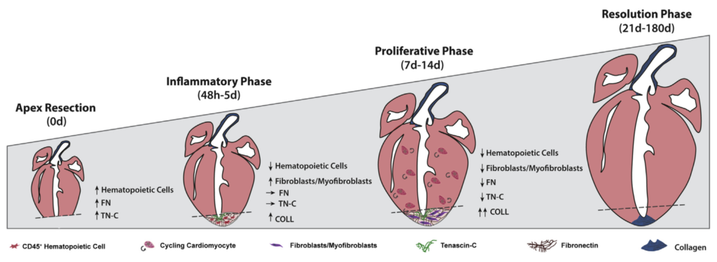
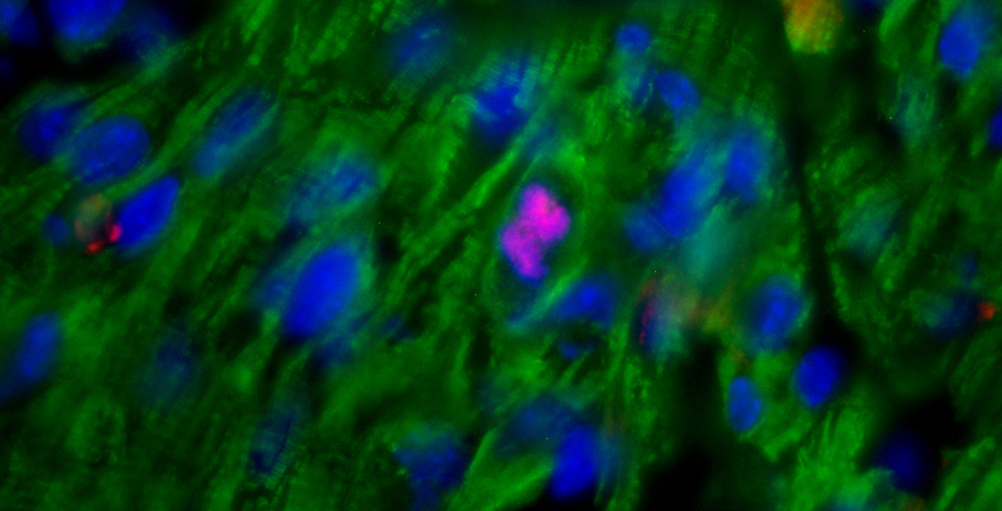
Abstract: So far, opposing outcomes have been reported following neonatal apex resection in mice, questioning the validity of this injury model to investigate regenerative mechanisms. We performed a systematic evaluation, up to 180 days after surgery, of the pathophysiological events activated upon apex resection. In response to cardiac injury, we observed increased cardiomyocyte proliferation in remote and apex regions, neovascularization, and local fibrosis. In adulthood, resected hearts remain consistently shorter and display permanent fibrotic tissue deposition in the center of the resection plane, indicating limited apex regrowth. However, thickening of the left ventricle wall, explained by an upsurge in cardiomyocyte proliferation during the initial response to injury, compensated cardiomyocyte loss and supported normal systolic function. Thus, apex resection triggers both regenerative and reparative mechanisms, endorsing this injury model for studies aimed at promoting cardiomyocyte proliferation and/or downplaying fibrosis.
Vai all’articolo originale (in inglese): Sampaio-Pinto V, Rodrigues S, Laúndos T, Silva E, Nóvoa F, Silva A, Cerqueira R, Resende T, Pianca N, Leite-Moreira A, D’Uva G, Thorsteinsdóttir S, Pinto-do-Ó P, Santos Nascimento D. Neonatal Apex Resection Triggers Cardiomyocyte Proliferation, Neovascularization and Functional Recovery Despite Local Fibrosis. Stem Cell Report, 2018
