BMP7 as a Key to Heart Regeneration: Our Progress

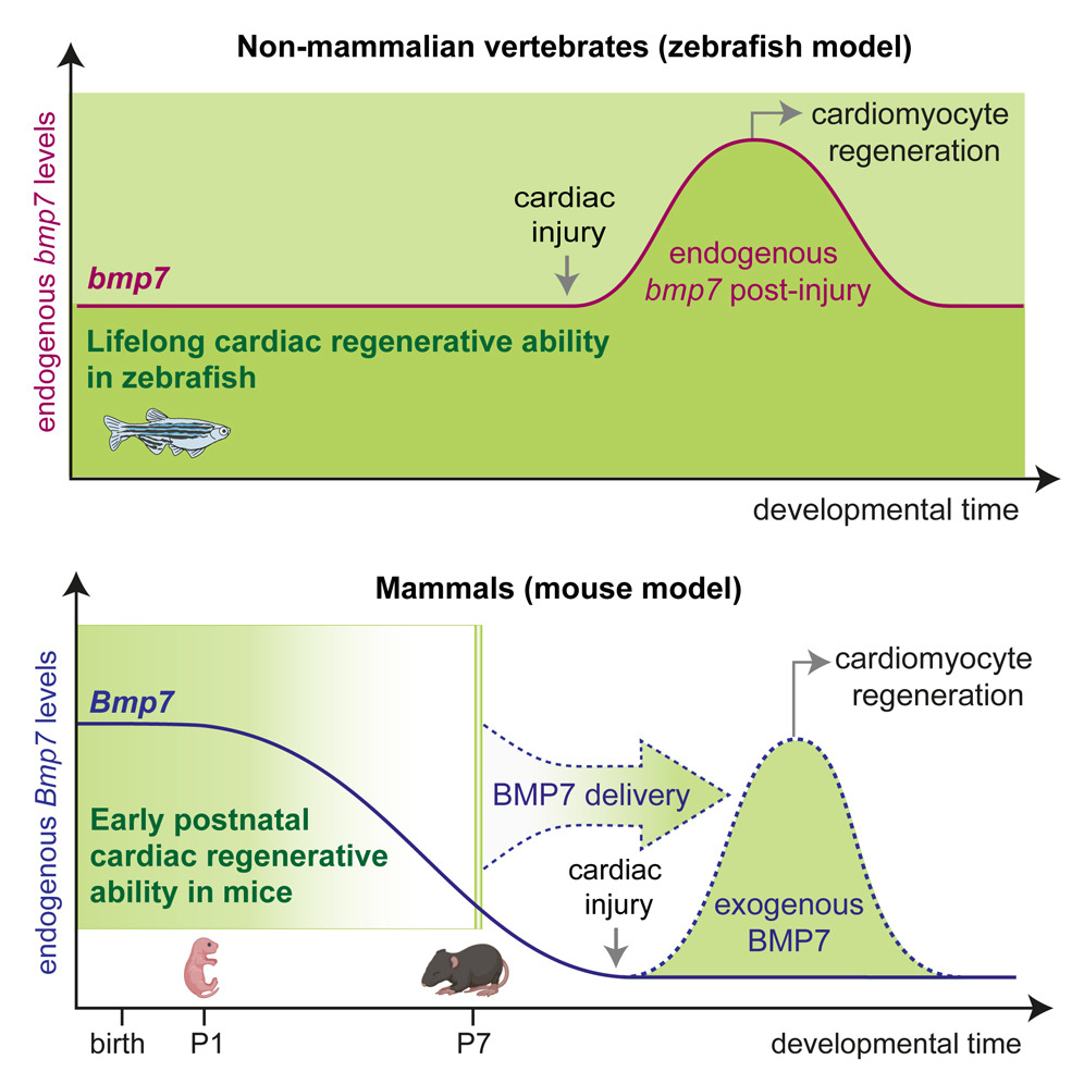
We are thrilled to announce our latest publication in Cell Reports! Our research indicates that a decrease in the production of growth factors, particularly BMP7, during early postnatal development contributes to the loss of regenerative capacity of the mammalian heart.
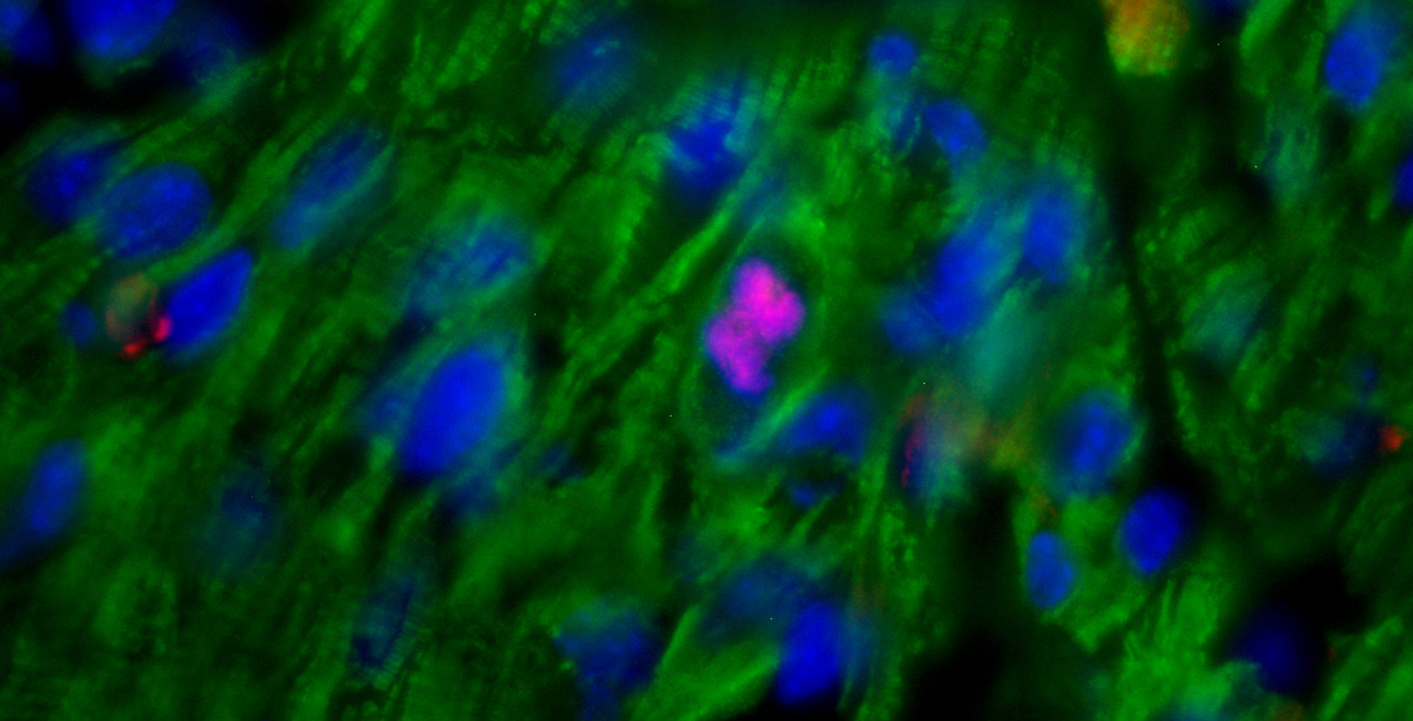
We demonstrate that BMP7 supports cardiomyocyte proliferation during the neonatal stage, and its administration boosts cardiomyocyte proliferation in postnatal life, even in adulthood, and especially after myocardial infarction. These findings suggest that BMP7 administration holds promise as a therapeutic approach for heart regeneration. Moreover, our study finds support from the zebrafish model, which naturally regenerates injured hearts. In this regard, inhibiting BMP7 impeded cardiomyocyte regeneration post-cardiac injury, while its induction accelerated the process. We believe our findings pave the way for heart regenerative therapies based on the administration of BMP7.
Congratulations to Chiara Bongiovanni for leading the project, and congratulations to the other lab team members Irene Del Bono, Carmen Miano, Stefano Boriati, Silvia Da Pra, Francesca Sacchi, Francesca Pontis, and Ilaria Petraroia for their help and support in experimental activities. We express our gratitude to collaborators who were instrumental in the success of this project, particularly the research groups led by Eldad Tzahor(Weizmann Institute of Science, Israel), Gilbert Weidinger (University of Ulm, Germany), Stephan Heermann (University of Friburg, Germany), Mattia Lauriola, and Carlo Ventura (University of Bologna, Italy).

Original Article Link (open access): https://dx.doi.org/10.1016/j.celrep.2024.114162
Our review on cardiotoxicity of anticancer therapies is online!

Unfortunately, anticancer drugs can induce side effects on heart tissue. This phenomenon, known as cardiotoxicity, in severe cases, can reduce the quality and life expectancy of cancer patients, regardless of the oncological prognosis.

The good news is that the complex molecular mechanisms responsible for these effects in different classes of anticancer drugs are beginning to emerge, and as a result, potential therapeutic approaches to protect the heart from these effects are beginning to be proposed.

#cardiotossicità #farmaciantitumorali #ricercascientifica #medicina #salute #unibo #irccsmultimedica #conFondazioneCariplo


Abstract: Chemotherapy and targeted therapies have significantly improved the prognosis of oncology patients. However, these antineoplastic treatments may also induce adverse cardiovascular effects, which may lead to acute or delayed onset of cardiac dysfunction. These common cardiovascular complications, commonly referred to as cardiotoxicity, not only may require the modification, suspension, or withdrawal of life-saving antineoplastic therapies, with the risk of reducing their efficacy, but can also strongly impact the quality of life and overall survival, regardless of the oncological prognosis. The onset of cardiotoxicity may depend on the class, dose, route, and duration of administration of anticancer drugs, as well as on individual risk factors. Importantly, the cardiotoxic side effects may be reversible, if cardiac function is restored upon discontinuation of the therapy, or irreversible, characterized by injury and loss of cardiac muscle cells. Subclinical myocardial dysfunction induced by anticancer therapies may also subsequently evolve in symptomatic congestive heart failure. Hence, there is an urgent need for cardioprotective therapies to reduce the clinical and subclinical cardiotoxicity onset and progression and to limit the acute or chronic manifestation of cardiac damages. In this review, we summarize the knowledge regarding the cellular and molecular mechanisms contributing to the onset of cardiotoxicity associated with common classes of chemotherapy and targeted therapy drugs. Furthermore, we describe and discuss current and potential strategies to cope with the cardiotoxic side effects as well as cardioprotective preventive approaches that may be useful to flank anticancer therapies.

Go to the original article: Morelli MB, Miano C, Bongiovanni C, Sacchi F, Da Pra S, Lauriola M and D’Uva G. Cardiotoxicity of Anticancer Drugs: Molecular Mechanisms and Strategies for Cardioprotection. Frontiers in Cardiovascular Medicine, 2022

Our publication (review) on “direct cardiogenesis” strategies for heart regeneration is online!

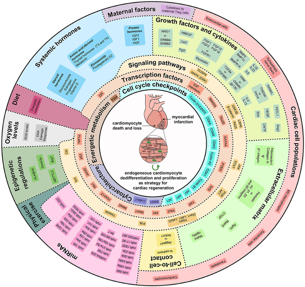
Is it possible to reawaken the intrinsic cardiac regenerative potential after major injuries, for example induced by myocardial infarction? We are happy to share our publication (review) on an emerging, promising and rapidly evolving approach for heart regeneration based on stimulating the proliferation of endogenous cardiac muscle cells.

Abstract:  Despite considerable efforts carried out to develop stem/progenitor cell-based technologies aiming at replacing and restoring the cardiac tissue following severe damages, thus far no strategies based on adult stem cell transplantation have been demonstrated to efficiently generate new cardiac muscle cells. Intriguingly, dedifferentiation, and proliferation of pre-existing cardiomyocytes and not stem cell differentiation represent the preponderant cellular mechanism by which lower vertebrates spontaneously regenerate the injured heart. Mammals can also regenerate their heart up to the early neonatal period, even in this case by activating the proliferation of endogenous cardiomyocytes. However, the mammalian cardiac regenerative potential is dramatically reduced soon after birth, when most cardiomyocytes exit from the cell cycle, undergo further maturation, and continue to grow in size. Although a slow rate of cardiomyocyte turnover has also been documented in adult mammals, both in mice and humans, this is not enough to sustain a robust regenerative process. Nevertheless, these remarkable findings opened the door to a branch of novel regenerative approaches aiming at reactivating the endogenous cardiac regenerative potential by triggering a partial dedifferentiation process and cell cycle re-entry in endogenous cardiomyocytes. Several adaptations from intrauterine to extrauterine life starting at birth and continuing in the immediate neonatal period concur to the loss of the mammalian cardiac regenerative ability. A wide range of systemic and microenvironmental factors or cell-intrinsic molecular players proved to regulate cardiomyocyte proliferation and their manipulation has been explored as a therapeutic strategy to boost cardiac function after injuries. We here review the scientific knowledge gained thus far in this novel and flourishing field of research, elucidating the key biological and molecular mechanisms whose modulation may represent a viable approach for regenerating the human damaged myocardium.

Go to the full article: : Bongiovanni C, Sacchi F, Da Pra S, Pantano E, Miano C, Morelli MB and D’Uva G. Reawakening the intrinsic cardiac regenerative potential: molecular strategies to boost dedifferentiation and proliferation of endogenous cardiomyocytes. Frontiers in Cardiovascular Medicine – Cardiovascular Biologics and Regenerative Medicine, 2021

#research #heart #regeneration #cardiomyocyte #proliferation #myocardialinfarction #duvalab

Thanks to AIRC funding we have the opportunity to implement our research in the cancer field and to recruit two researchers in our group

We have recently received wonderful news: one of the funding requests has gone through! Indeed, an AIRC grant will allow us to implement our research on cancer, in parallel to ongoing studies in the field of cardiac regeneration and cardiotoxicity of anticancer therapies.

In fact, our laboratory aims at understanding the molecular mechanisms underlying cell differentiation and proliferation to develop new strategies to “block” cancer cells and to “unlock” the cardiac regenerative potential. In particular, with this project, we aim at studying the deregulations responsible for the development and progression of basal-like breast cancer, an aggressive tumor subtype.

In this research project, we will analyze the role and the molecular mechanisms activated by growth factors in cell differentiation. We hypothesize that pushing basal breast cancer cells towards a more differentiated phenotype would reduce their aggressiveness. The project has the potential to develop new differentiation strategies to prevent primary tumor cells from disseminating throughout the body and creating the so-called metastases, one of the major problems in solid tumor progression.

This funding will give us the opportunity to recruit two researchers (postgraduate or postdoc) in our research group in Bologna, from February / March 2021 for a total of five-year. Pre-doctoral researchers may also have the opportunity to start a PhD track. Interested and motivated researchers can contact us by email (duva.gabriele@gmail.com), attaching their CV.

Our researcher Dr. Nicola Pianca is the winner of the International Society for Heart Reseach Fellowship 2019!

Congratulations to Nicola Pianca, a postdoc researcher of our team, winner of the annual fellowship offered by SERVIER in collaboration with the European Section of the International Society for Heart Reseach (ISHR)

Dr. Pianca’s project focuses on the interaction between growth factors and hormones in the context of heart regeneration. Preliminary data are very promising and have already been presented at various international conferences over the last year, arousing some interest in the international scientific community.

In June 2019, we will present the project at the World Congress of the International Society for Heart Research (ISHR).

Our article on heart development is out in Nature!

Glad we could be part of this international project on heart development! Congratulations to Gonzalo del Monte Nieto and Richard Harvey!

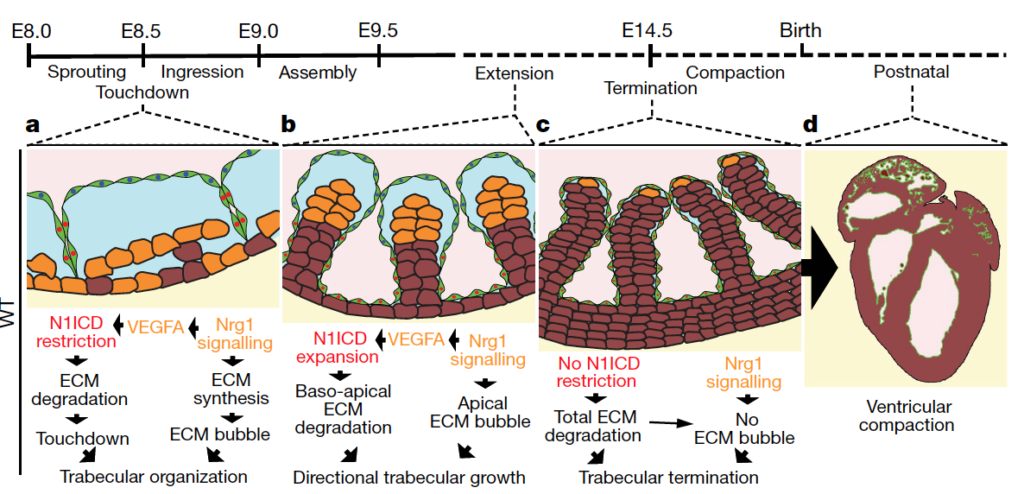
Abstract: In vertebrate hearts, the ventricular trabecular myocardium develops as a sponge-like network of cardiomyocytes that is critical for contraction and conduction, ventricular septation, papillary muscle formation and wall thickening through the process of compaction. Defective trabeculation leads to embryonic lethality or non-compaction cardiomyopathy (NCC). There are divergent views on when and how trabeculation is initiated in different species. In zebrafish, trabecular cardiomyocytes extrude from compact myocardium, whereas in chicks, chamber wall thickening occurs before overt trabeculation. In mice, the onset of trabeculation has not been described, but is proposed to begin at embryonic day 9.0, when cardiomyocytes form radially oriented ribs. Endocardium–myocardium communication is essential for trabeculation, and numerous signalling pathways have been identified, including Notch and Neuregulin (NRG). Late disruption of the Notch pathway causes NCC5. Whereas it has been shown that mutations in the extracellular matrix (ECM) genes Has2 and Vcan prevent the formation of trabeculae in mice and the matrix metalloprotease ADAMTS1 promotes trabecular termination, the pathways involved in ECM dynamics and the molecular regulation of trabeculation during its early phases remain unexplored. Here we present a model of trabeculation in mice that integrates dynamic endocardial and myocardial cell behaviours and ECM remodelling, and reveal new epistatic relationships between the involved signalling pathways. NOTCH1 signalling promotes ECM degradation during the formation of endocardial projections that are critical for individualization of trabecular units, whereas NRG1 promotes myocardial ECM synthesis, which is necessary for trabecular rearrangement and growth. These systems interconnect through NRG1 control of Vegfa, but act antagonistically to establish trabecular architecture. These insights enabled the prediction of persistent ECM and cardiomyocyte growth in a mouse NCC model, providing new insights into the pathophysiology of congenital heart disease.

Go to the full article: Del Monte-Nieto G, Ramialison M, Adam AAS, Wu B, Aharonov A, D’Uva G, Bourke LM, Pitulescu ME, Chen H, de la Pompa JL, Shou W, Adams RH, Harten SK, Tzahor E, Zhou B and Harvey RP. Control of cardiac jelly dynamics by NOTCH1 and NRG1 defines the building plan for trabeculation. Nature, 2018

Fellowship opportunity in our lab

We are recruiting a research fellow at MultiMedica ONLUS Foundation (Milan). The contract is for 1 year, starting from June 2018, and potentially renewable for a total of three years.
The project is funded by Cariplo Foundation and the research will be conducted in our laboratory, which forms a bridge between the fields of cancer, cardiotoxicity of anticancer therapies and cardiac regeneration.

The selected research fellow will develop a project on the cardiotoxicity of anti-cancer therapies. Cardio-toxicity is a common side effect of chemotherapy and targeted anti-cancer therapies, strongly impacting on the quality of life and the overall survival, regardless of the oncological prognosis. The goal is to develop strategies for reducing the cardiotoxic side effects of current anti-cancer therapies, while simultaneously improving their efficacy. By employing both breast cancer cells and primary mouse cardiomyocytes cultures in vitro, the research activities will be focused on the role of specific growth factors and receptors on cell differentiation status, and the impact on tumour growth and cardiotoxic side effects. In vivo analysis on mouse models will also be conducted.

Applicants requirements: Applicants must have a molecular-, cell- and biochemistry backgrounds, such as a Master Degree in Biotechnology, Biology or related field. Candidates must have at least one year of experience in a research laboratory. Familiarity with standard molecular biology techniques (Real Time PCR, Western Blot, Immunofluorescence analysis…), as well as the expertise in cardiac and/or tumour biology, are required. Passion for science as well as a positive attitude towards solving problems is required in order to pursue technically challenging and intellectually stimulating projects in the lab.

Other criteria: PhD Degree is not required, but is positively evaluated. Expertise in procedures for the induction of cardiac damage (by cardiotoxic drugs or myocardial infarction) as well as in the analysis of microarray o RNA-seq datasets is a plus factor for the selection process.

How to apply: Applicants should send their CV by email with subject line “Research fellowships at Fondazione MultiMedica ONLUS” to Dr. Gabriele D’Uva (gabriele.duva@multimedica.it). One or more letters of recommendation are welcome, although not strictly required. Interviews will be conducted for selected candidates until the position is filled.

Previous publications relevant to the project:
• D’Uva G et al., ERBB2 triggers mammalian heart regeneration by promoting cardiomyocyte dedifferentiation and proliferation. Nature Cell Biology, 2015
• D’Uva G and Tzahor E, The key roles of ERBB2 in cardiac regeneration. Cell Cycle. 2015
• Yutzey KE. Regenerative biology: Neuregulin 1 makes heart muscle. Nature, 2015 (News & Views on our article)
• D’Uva G and Lauriola M, Towards the emerging cross-talk: ERBB family and steroid hormones. Seminars in Cell and Developmental Biology, 2016

Our article on heart regeneration is out in Stem Cell Reports!

Happy to have collaborated to this international project on mammalian heart regeneration! Congratulations to Diana Nascimento and her team!

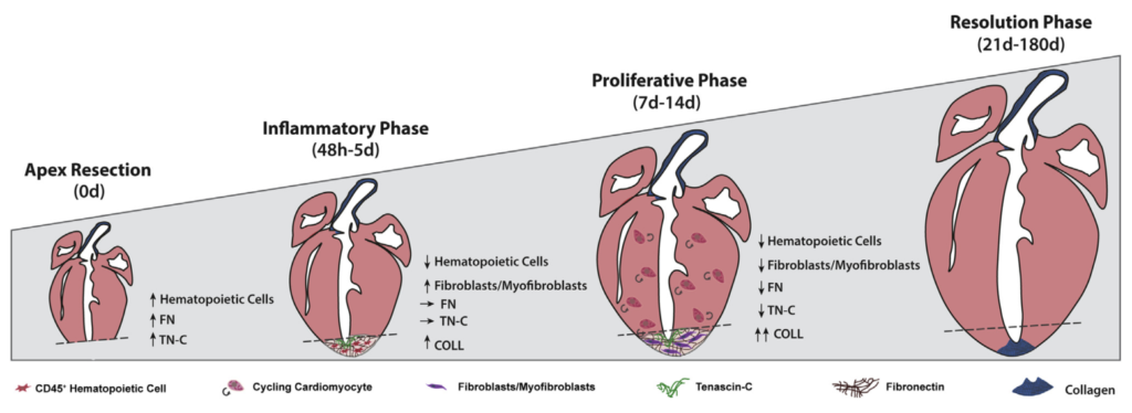
Abstract: So far, opposing outcomes have been reported following neonatal apex resection in mice, questioning the validity of this injury model to investigate regenerative mechanisms. We performed a systematic evaluation, up to 180 days after surgery, of the pathophysiological events activated upon apex resection. In response to cardiac injury, we observed increased cardiomyocyte proliferation in remote and apex regions, neovascularization, and local fibrosis. In adulthood, resected hearts remain consistently shorter and display permanent fibrotic tissue deposition in the center of the resection plane, indicating limited apex regrowth. However, thickening of the left ventricle wall, explained by an upsurge in cardiomyocyte proliferation during the initial response to injury, compensated cardiomyocyte loss and supported normal systolic function. Thus, apex resection triggers both regenerative and reparative mechanisms, endorsing this injury model for studies aimed at promoting cardiomyocyte proliferation and/or downplaying fibrosis.

Go to the full article: Sampaio-Pinto V, Rodrigues S, Laúndos T, Silva E, Nóvoa F, Silva A, Cerqueira R, Resende T, Pianca N, Leite-Moreira A, D’Uva G, Thorsteinsdóttir S, Pinto-do-Ó P, Nascimento DS. Neonatal Apex Resection Triggers Cardiomyocyte Proliferation, Neovascularization and Functional Recovery Despite Local Fibrosis. Stem Cell Report, 2018

D’Uva lab receives Cariplo grant award on cancer and cardiotoxicity

We have just received very good news: we won the research grant “Cariplo – Young Researchers“.
This grant will allow us, in parallel to ongoing studies in the field of cardiac regeneration following a heart attack, to expand our research on cancer and the emerging problem of cardiotoxicity of anticancer therapies.
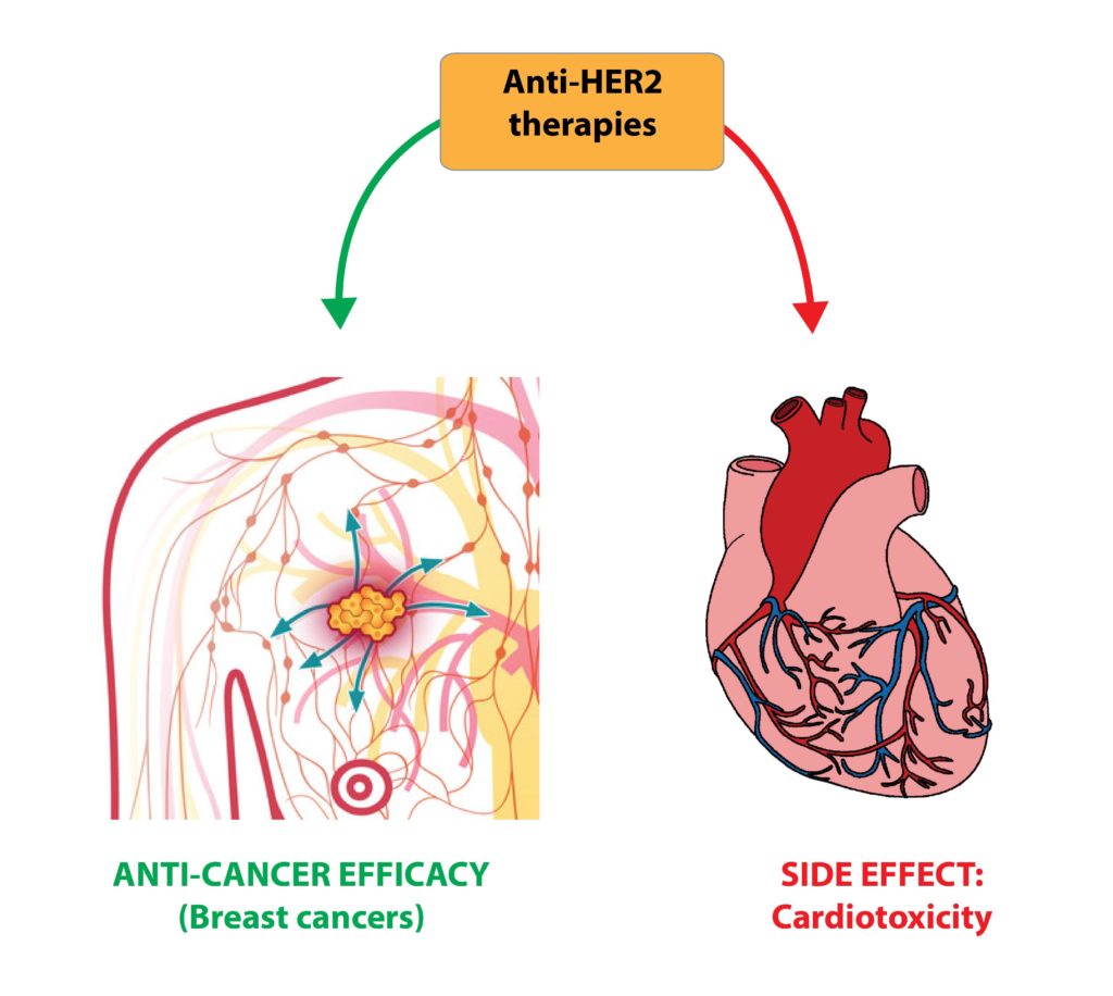
Cardiotoxicity, i.e. toxic effects on heart function, is responsible for a poor quality of life and reduced survival of cancer patients, regardless of the oncologic prognosis. The aim of our research is to develop innovative strategies to limit these side effects while increasing the efficacy of anti-cancer therapies targeting HER2 (a well-known oncogene also known as ERBB2) in breast cancer patients.
The project is in collaboration with the University of Bologna and Biomedical Research Foundation / University of Turin.
These funds will also give us the opportunity to recruit another researcher into my team. Soon we will post the details.
Happy 2018 to everyone!