Our article is out in Nature Communications! Happy to have collaborated to this ambitious international project on steroid hormones in cancer development!

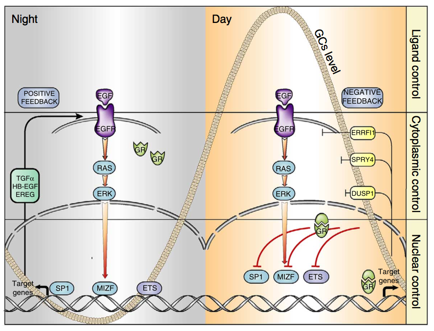
Signal transduction by receptor tyrosine kinases (RTKs) and nuclear receptors for steroid hormones is essential for body homeostasis, but the cross-talk between these receptor families is poorly understood. We observed that glucocorticoids inhibit signalling downstream of EGFR, an RTK. The underlying mechanism entails suppression of EGFR’s positive feedback loops and simultaneous triggering of negative feedback loops that normally restrain EGFR. Our studies in mice reveal that the regulation of EGFR’s feedback loops by glucocorticoids translates to circadian control of EGFR signalling: EGFR signals are suppressed by high glucocorticoids during the active phase (night-time in rodents), while EGFR signals are enhanced during the resting phase. Consistent with this pattern, treatment of animals bearing EGFR-driven tumours with a specific kinase inhibitor was more effective if administered during the resting phase of the day, when glucocorticoids are low. These findings support a circadian clock-based paradigm in cancer therapy.
Go to the full article: Mattia Lauriola, Yehoshua Enuka, Amit Zeisel, Gabriele D’Uva, Lee Roth, Michal Sharon-Sevilla, Moshit Lindzen, Kirti Sharma, Nava Nevo, Morris Feldman, Silvia Carvalho, Hadas Cohen-Dvashi, Merav Kedmi, Nir Ben-Chetrit, Alon Chen, Rossella Solmi, Stefan Wiemann, Fernando Schmitt, Eytan Domany & Yosef Yarden. Diurnal suppression of EGFR signalling by glucocorticoids and implications for tumour progression and treatment. Nature Communications, 2015


MEDIA COVERAGE

06.10.2014 – Science Daily Tumors might grow faster at nigh


06.10.2014 – NATURE WORLD NEWS: Is Cancer Growth Nocturnal?


06.10.2014 – Softpedia: Cancer Tumors Appear to Grow Faster and Spread More Easily at Night


06.10.2014 – Medical Xpress: Tumors might grow faster at night: Hormone that keeps us alert also suppresses the spread of cancer


06.10.2014 – Senior Journal: Cancer Grows at Night, Maybe That’s When to Attack, New Study Says


07.10.2014 – Daily Mail: Cancerous tumours ‘grow faster at night’ – and drugs to fight the disease might work better during sleep, study finds


07.10.2014 – Counsel&Heal: Cancer Treatment More Efficient During Night Time:Study


07.10.2014 – Science 2.0: Cancer Might Grow Faster At Night


07.10.2014 – The Health Site: Revealed — cancer spreads during nights


07.10.2014 – bhataramedia: Tumor Dapat Tumbuh Lebih Cepat Di Malam Hari


07.10.2014 – Jews News: Israeli study: Tumors might grow more quickly at night


08.10.2014 – The Times of Israel: Tumors may grow faster at night, Israeli study shows


08.10.2014 – Journal de la Science: Les tumeurs grossiraient plus vite la nuit


08.10.2014 – Haaretz: Night time may be the right time to treat cancer, find Israeli scientists


09.10.2014 – Weizmann USA: Why Cancer Drugs May Work Better While You Sleep


09.10.2014 – Nature子刊:夜间癌细胞扩散的更快?


09.10.2014 – TIME: Why Cancer Drugs May Work Better While You Sleep


09-10-2014 – Medisite: CANCER : LES MÉDICAMENTS PLUS EFFICACES LA NUIT


10.10.2014 – TopSante: Cancer : les tumeurs se développent plus vite la nuit


10.10.2014 – Forbes: Cancer May Grow Faster While We Sleep


10.10.2014 – Jerusalem Post Tumors may grow faster at night, say Weizmann scientists


10.10.2014 – Medcenter: Una hormona que nos mantiene alerta también suprime la diseminación del cáncer


16.10.2014 – EACR (European Association for Cancer Research): TUMOURS MIGHT GROW FASTER AT NIGHT


16.10.2014 – Revista Genetica Medica: Ritmos circadianos y tratamiento contra el cáncer


17.10.2014 – University Herald: Hormone Active During Day Supresses Growth of Cancer Cells, Study


20.10.2014 – Stato Quotidiano: Ricercatrice Manfredonia: “tumori crescono più in fretta di notte”


24.10.2014 – Huffington Post: Does Cancer Grow More Aggressively at Night?
Our article on post-transcriptional mechanisms in cancer stem cells is out!

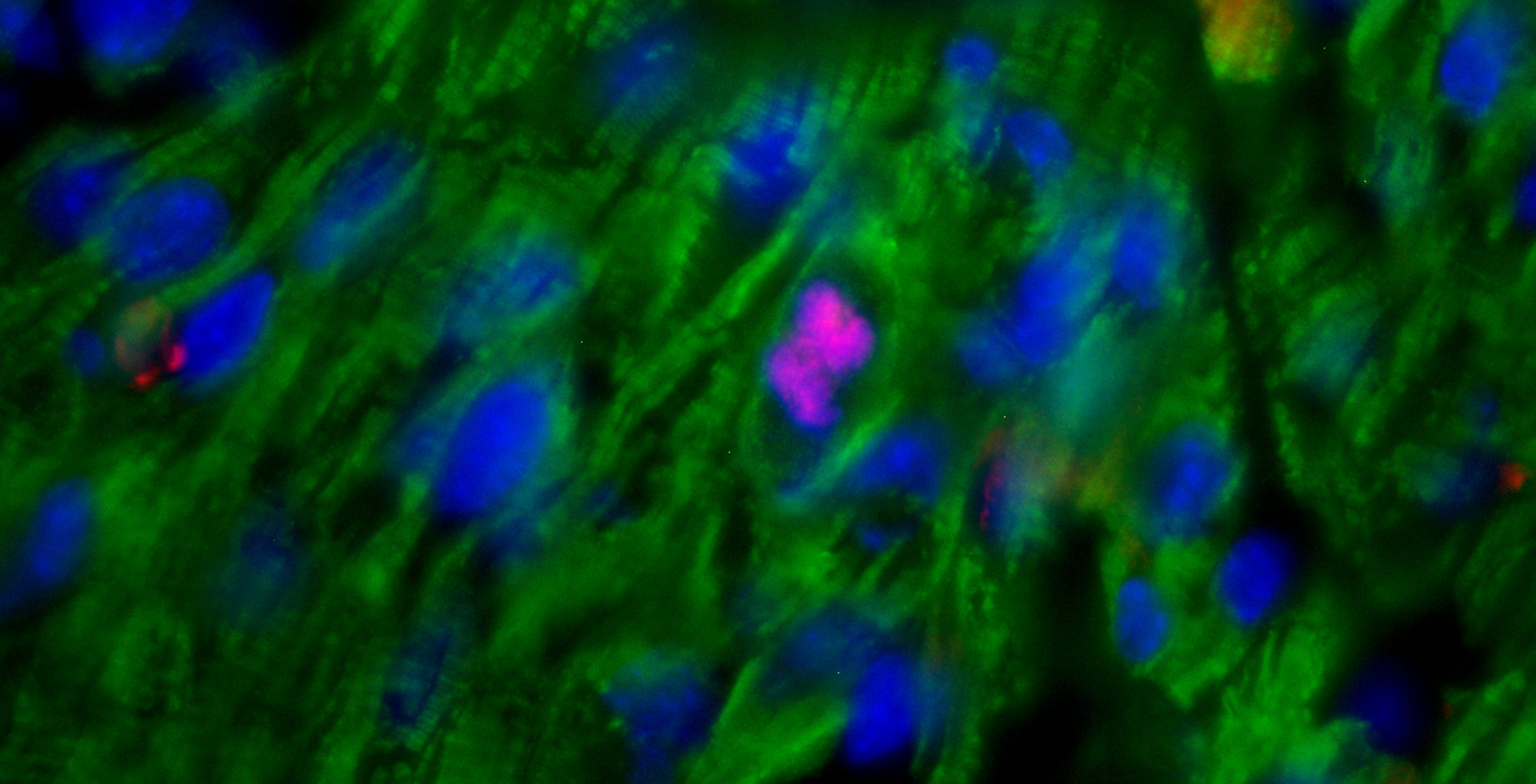
Hypoxia has been long-time acknowledged as major cancer-promoting microenvironment. In such an energy-restrictive condition, post-transcriptional mechanisms gain importance over the energy-expensive gene transcription machinery. Here we show that the onset of hypoxia-induced cancer stem cell features requires the beta-catenin-dependent post-transcriptional up-regulation of CA9 and SNAI2 gene expression. In response to hypoxia, beta-catenin moves from the plasma membrane to the cytoplasm where it binds and stabilizes SNAI2 and CA9 mRNAs, in cooperation with the mRNA stabilizing protein HuR. We also provide evidence that the post-transcriptional activity of cytoplasmic beta-catenin operates under normoxia in basal-like/triple-negative breast cancer cells, where the beta-catenin knockdown suppresses the stem cell phenotype in vitro and tumor growth in vivo. In such cells, we unravel the generalized involvement of the beta-catenin-driven machinery in the stabilization of EGF-induced mRNAs, including the cancer stem cell regulator IL6. Our study highlights the crucial role of post-transcriptional mechanisms in the maintenance/acquisition of cancer stem cell features and suggests that the hindrance of cytoplasmic beta-catenin function may represent an unprecedented strategy for targeting breast cancer stem/basal-like cells.

Go to the full article: Gabriele D’Uva*, Sara Bertoni, Mattia Lauriola, Sabrina De Carolis, Annalisa Pacilli, Laura D’Anello, Donatella Santini, Mario Taffurelli, Claudio Ceccarelli, Yosef Yarden, Lorenzo Montanaro, Massimiliano Bonafé, Gianluca Storci. Beta-Catenin/HuR Post-Transcriptional Machinery Governs Cancer Stem Cell Features in Response to Hypoxia. PloS One, 2013 (co-corresponding author)

Our article is out in JCI! Happy to have collaborated to this international project on physiological hematopoietic stem cell egress!

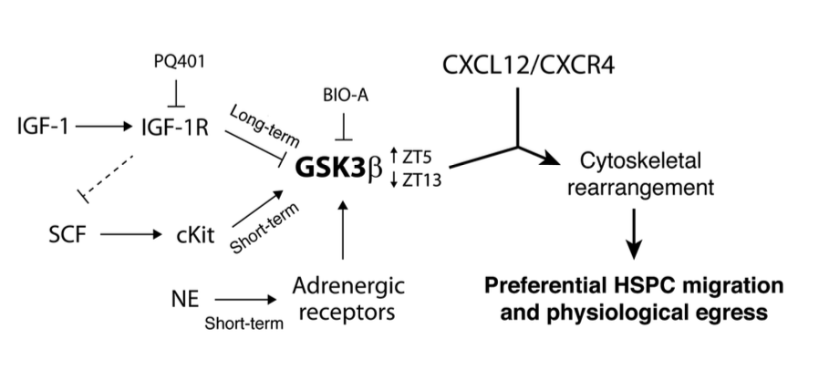
Regulation of hematopoietic stem and progenitor cell (HSPC) steady-state egress from the bone marrow (BM) to the circulation is poorly understood. While glycogen synthase kinase-3β (GSK3β) is known to participate in HSPC proliferation, we revealed an unexpected role in the preferential regulation of CXCL12-induced migration and steady-state egress of murine HSPCs, including long-term repopulating HSCs, over mature leukocytes. HSPC egress, regulated by circadian rhythms of CXCL12 and CXCR4 levels, correlated with dynamic expression of GSK3β in the BM. Nevertheless, GSK3β signaling was CXCL12/CXCR4 independent, suggesting that synchronization of both pathways is required for HSPC motility. Chemotaxis of HSPCs expressing higher levels of GSK3β compared with mature cells was selectively enhanced by stem cell factor-induced activation of GSK3β. Moreover, HSPC motility was regulated by norepinephrine and insulin-like growth factor-1 (IGF-1), which increased or reduced, respectively, GSK3β expression in BM HSPCs and their subsequent egress. Mechanistically, GSK3β signaling promoted preferential HSPC migration by regulating actin rearrangement and microtubuli turnover, including CXCL12-induced actin polarization and polymerization. Our study identifies a previously unknown role for GSK3β in physiological HSPC motility, dictating an active, rather than a passive, nature for homeostatic egress from the BM reservoir to the blood circulation.

Go to the full article: Kfir Lapid, Tomer Itkin, Gabriele D’Uva, Yossi Ovadya, Aya Ludin, Giulia Caglio, Alexander Kalinkovich, Karin Golan, Ziv Porat, Massimo Zollo, Tsvee Lapidot. GSK3β regulates physiological migration of stem/progenitor cells via cytoskeletal rearrangement. The Journal of clinical investigation, 2013

Our article is out in Nature Immunology! Happy to have collaborated to this international project on a new component of the hematopoietic stem cell niche!

Hematopoietic stem and progenitor cells (HSPCs) are regulated by various bone marrow stromal cell types. Here we identified rare activated bone marrow monocytes and macrophages with high expression of α-smooth muscle actin (α-SMA) and the cyclooxygenase COX-2 that were adjacent to primitive HSPCs. These myeloid cells resisted radiation-induced cell death and further upregulated COX-2 expression under stress conditions. COX-2-derived prostaglandin E(2) (PGE(2)) prevented HSPC exhaustion by limiting the production of reactive oxygen species (ROS) via inhibition of the kinase Akt and higher stromal-cell expression of the chemokine CXCL12, which is essential for stem-cell quiescence. Our study identifies a previously unknown subset of α-SMA(+) activated monocytes and macrophages that maintain HSPCs and protect them from exhaustion during alarm situations.

Go to the full article: Aya Ludin, Tomer Itkin, Shiri Gur-Cohen, Alexander Mildner, Elias Shezen, Karin Golan, Orit Kollet, Alexander Kalinkovich, Ziv Porat, Gabriele D’Uva, Amir Schajnovitz, Elena Voronov, David A Brenner, Ron N Apte, Steffen Jung, Tsvee Lapidot. Monocytes-macrophages that express α-smooth muscle actin preserve primitive hematopoietic cells in the bone marrow. Nature Immunology, 2012

Our article is out in Nature Immunology! Happy to have collaborated to this international project on the dynamic regulation of the hematopoietic stem cell niche!

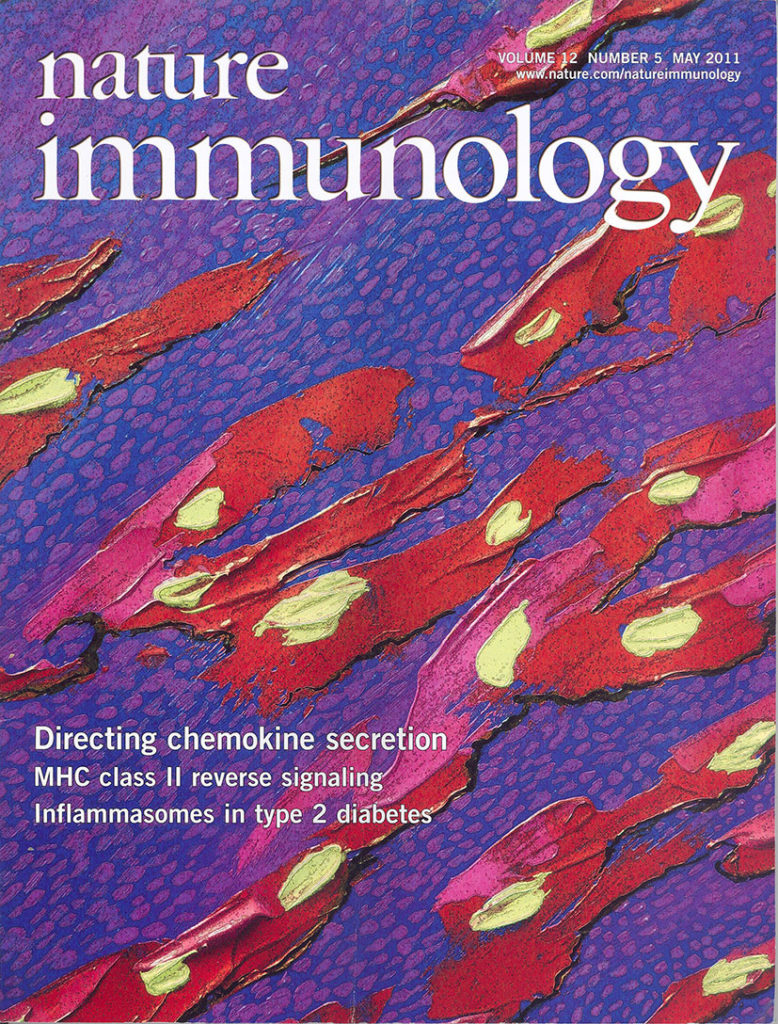
The chemokine CXCL12 is essential for the function of hematopoietic stem and progenitor cells. Here we report that secretion of functional CXCL12 from human bone marrow stromal cells (BMSCs) was a cell contact-dependent event mediated by connexin-43 (Cx43) and Cx45 gap junctions. Inhibition of connexin gap junctions impaired the secretion of CXCL12 and homing of leukocytes to mouse bone marrow. Purified human CD34(+) progenitor cells did not adhere to non contacting BMSCs, which led to a much smaller pool of immature cells. Calcium conduction activated signaling by cAMP-protein kinase A (PKA) and induced CXCL12 secretion mediated by the GTPase RalA. Cx43 and Cx45 additionally controlled Cxcl12 transcription by regulating the nuclear localization of the transcription factor Sp1. We suggest that BMSCs form a dynamic syncytium via connexin gap junctions that regulates CXCL12 secretion and the homeostasis of hematopoietic stem cells.

Go to the full article: Schajnovitz A, Itkin T, D’Uva G, Kalinkovich A, Golan K, Ludin A, Cohen D, Shulman Z, Avigdor A, Nagler A, Kollet O, Seger R, Lapidot T. CXCL12 secretion by bone marrow stromal cells is dependent on cell contact and mediated by connexin-43 and connexin-45 gap junctions. Nature Immunology, 2011

This article has been been selected for the cover of of Nature Immunology (May 2011) and commented in Nature Immunology – News and Views 

Happy to have collaborated to this project on inflammation and epigenetic regulations!

BACKGROUND: Basal-like carcinoma are aggressive breast cancers that frequently carry p53 inactivating mutations, lack estrogen receptor-α (ERα) and express the cancer stem cell markers CD133 and CD44. These tumors also over-express Interleukin 6 (IL-6), a pro-inflammatory cytokine that stimulates the growth of breast cancer stem/progenitor cells.

RESULTS: Here we show that p53 deficiency in breast cancer cells induces a loss of methylation at IL-6 proximal promoter region, which is maintained by an IL-6 autocrine loop. IL-6 also elicits the loss of methylation at the CD133 promoter region 1 and of CD44 proximal promoter, enhancing CD133 and CD44 gene transcription. In parallel, IL-6 induces the methylation of estrogen receptor (ERα) promoter and the loss of ERα mRNA expression. Finally, IL-6 induces the methylation of IL-6 distal promoter and of CD133 promoter region 2, which harbour putative repressor regions.

CONCLUSION: We conclude that IL-6, whose methylation-dependent autocrine loop is triggered by the inactivation of p53, induces an epigenetic reprogramming that drives breast carcinoma cells towards a basal-like/stem cell-like gene expression profile.

Go to the full article: D’Anello L, Sansone P, Storci G, Mitrugno V, D’Uva G, Chieco P, Bonafé M. Epigenetic control of the basal-like gene expression profile via Interleukin-6 in breast cancer cells. Molecular Cancer, 2010

Happy to have collaborated to this project on inflammation and stem cells!

Extracellular and intracellular mediators of inflammation, such as tumor necrosis factor alpha (TNFα) and NF-kappaB (NF-κB), play major roles in breast cancer pathogenesis, progression and relapse. SLUG, a mediator of the epithelial-mesenchymal transition process, is over-expressed in CD44(+)/CD24(-) tumor initiating breast cancer cells and in basal-like carcinoma, a subtype of aggressive breast cancer endowed with a stem cell-like gene expression profile. Cancer stem cells also over-express members of the pro-inflammatory NF-κB network, but their functional relationship with SLUG expression in breast cancer cells remains unclear. Here, we show that TNFα treatment of human breast cancer cells up-regulates SLUG with a dependency on canonical NF-κB/HIF1α signaling, which is strongly enhanced by p53 inactivation. Moreover, SLUG up-regulation engenders breast cancer cells with stem cell-like properties including enhanced expression of CD44 and Jagged-1 in conjunction with estrogen receptor alpha down-regulation, growth as mammospheres, and extracellular matrix invasiveness. Our results reveal a molecular mechanism whereby TNFα, a major pro-inflammatory cytokine, imparts breast cancer cells with stem cell-like features, which are connected to increased tumor aggressiveness.

Go to the full article: Storci G, Sansone P, Mari S, D’Uva G, Tavolari S, Guarnieri T, Taffurelli M, Ceccarelli C, Santini D, Chieco P, Marcu KB, Bonafè M. TNFalpha up‐regulates SLUG via the NF‐kappaB/HIF1alpha axis, which imparts breast cancer cells with a stem cell‐like phenotype. J Cell Physiology, 2010